
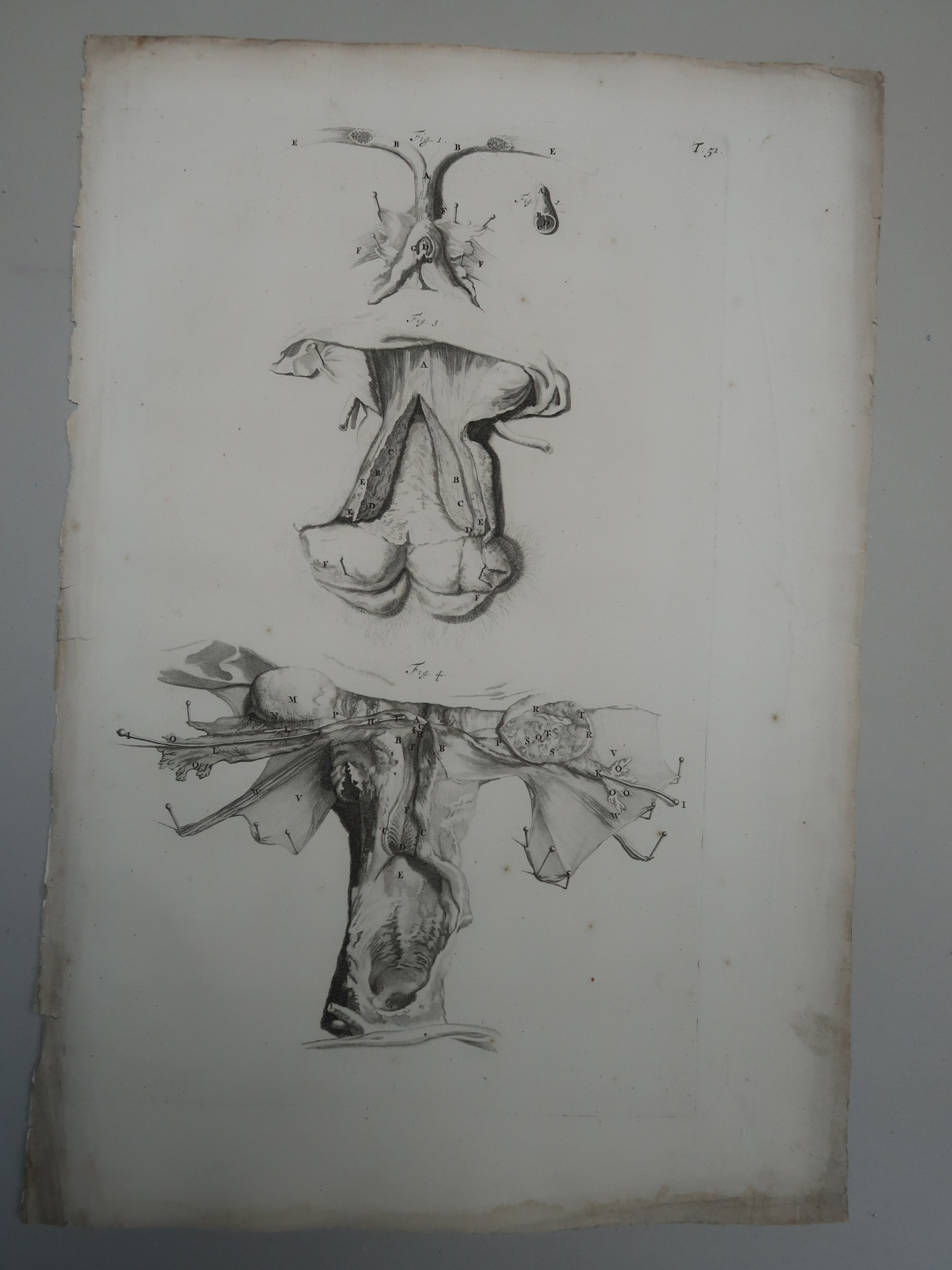
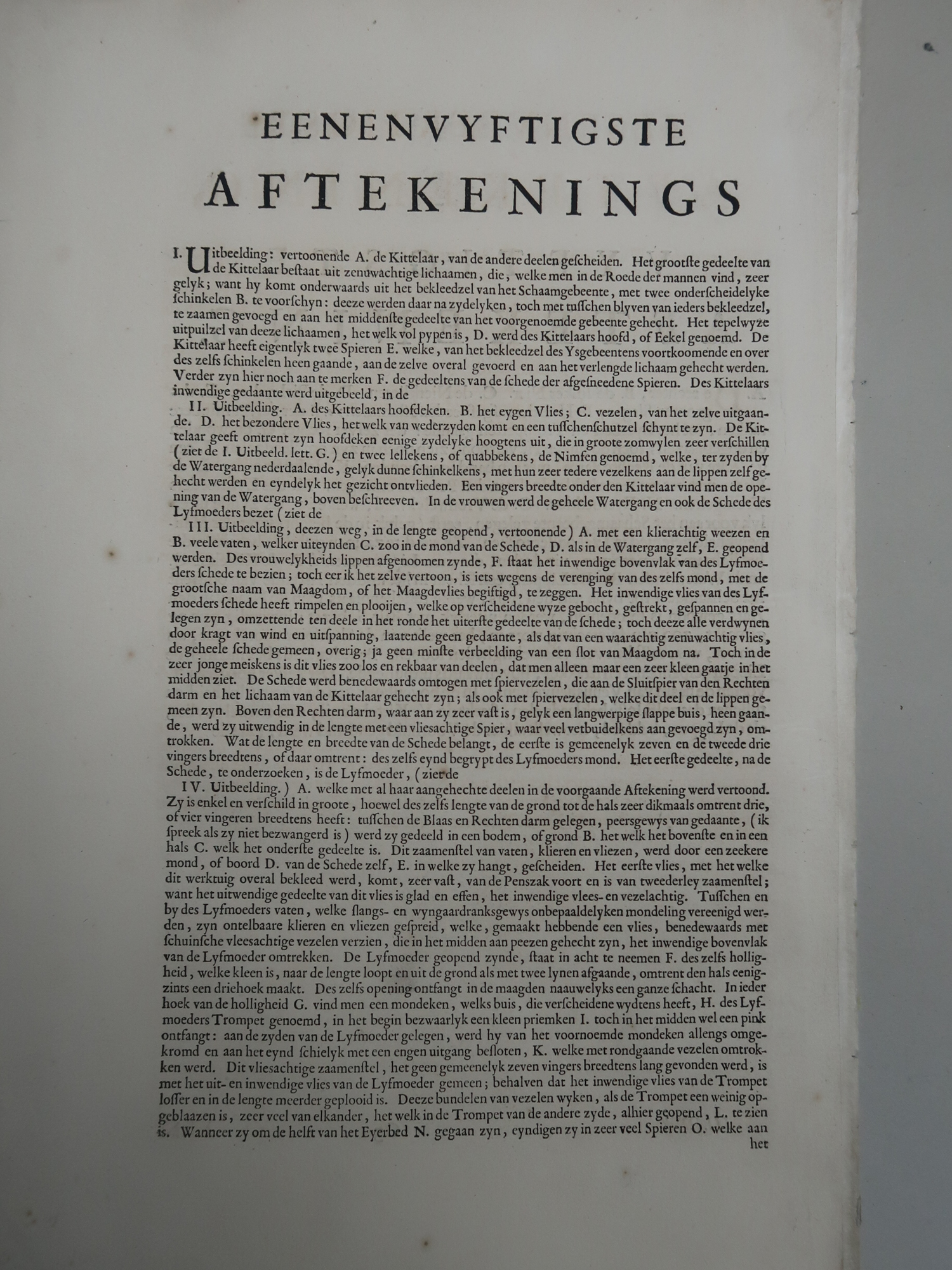
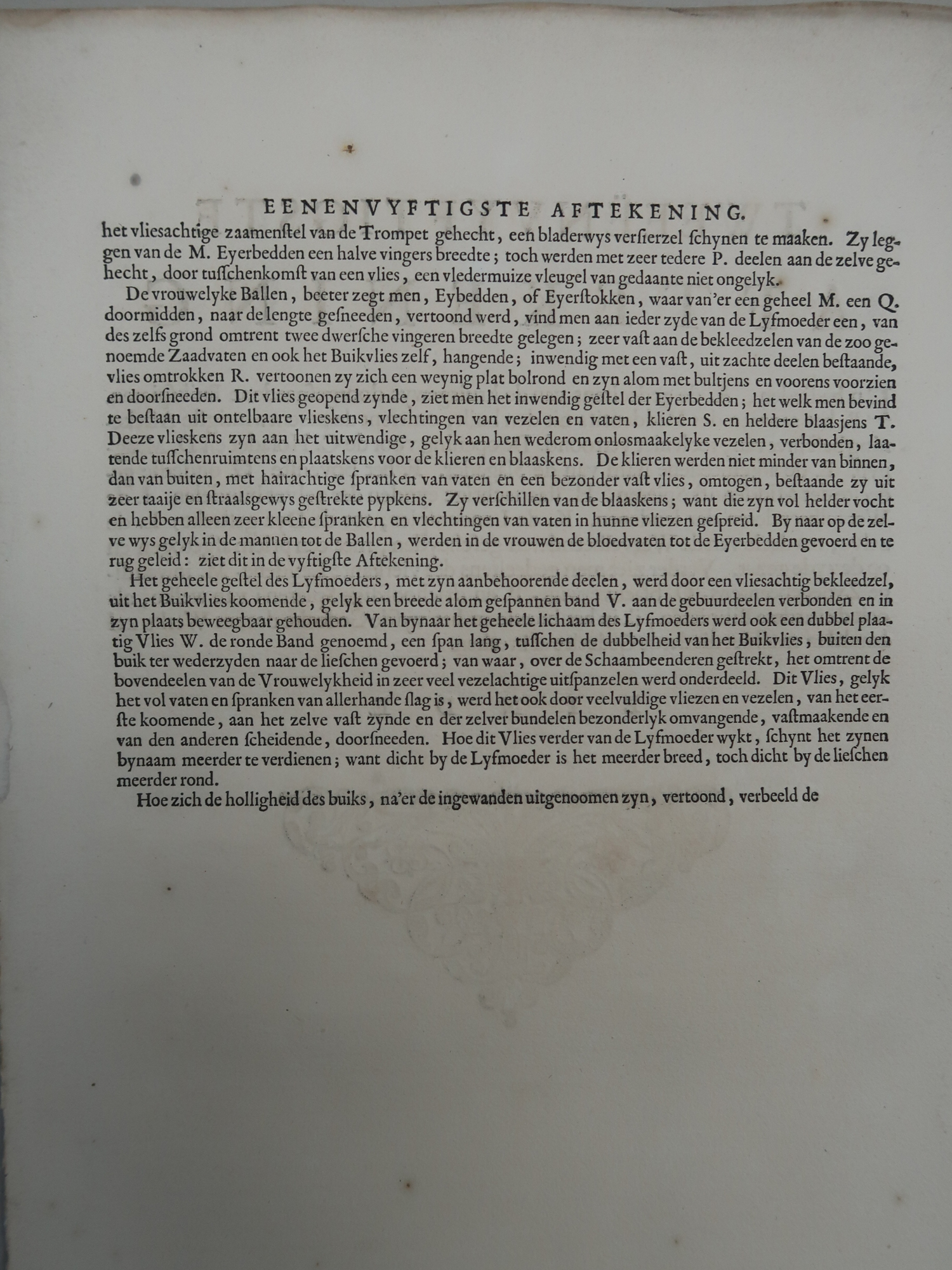
Description
Medium: Copperplate engraving on hand laid (verge) paper.
Sheet size: 34.5 x 51 cm (13.58 x 20.08 inch). Image size: 26 x 42 cm. (10.24 x 16.54 inch).
Condition: good, given age. Light foxing, creasing and soiling, mostly affecting the margin. Some tears in paper edges, not effecting the image. General age-related toning and/or occasional minor defects from handling. Please study scan carefully.
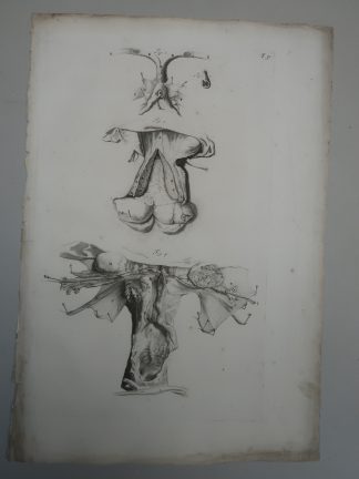
T.51.-ANATOMY-FEMALE-REPRODUCTIVE-ORGANS | BOEK-BIDLOO
BACKGROUND INFORMATION
Ontleding des Menschelyken Lichaams’, Utrecht, 1728, published by Jacob van Poolsum. A later Dutch version of Govard Bidloo’s most famous work, his monumental Anatomia humani corporis published in Amsterdam in 1685, containing 107 copperplate engravings. Like so many large and expensive anatomical atlases of the time, the work was not a financial success, and in 1690 he published a Dutch translation entitled, Ontleding des menschelyken lichaams, using the same plates. When this edition did not sell well either, Bidloo’s publisher sold 300 of the extra printed plates to William Cowper, a noted English anatomist. Cowper published the plates with his own, English language text in Oxford in 1698 under the title, Anatomy of the humane bodies, without mentioning Bidloo or the artists of the original plates. Cowper went so far as to use Bidloo’s engraved allegorical title page, amended with an irregular piece of paper lettered: “The anatomy of the humane bodies …,” which fits over the Dutch title (see a comparison here). A number of vitriolic exchanges took place between Bidloo and Cowper, including several pamphlets published in each anatomist’s defense. Cowper claimed, without much evidence presented, that the plates were not Bidloo’s at all, but that they were commissioned by Jan Swammerdam (1637?1680) and that after his death Swammerdam’s widow had sold them to Bidloo. The illustrations in Bidloos’ work were drawn by Gerard de Lairesse (1640?1711) and engraved by Abraham Blooteling (1640?1690) and Peter van Gunst (1659??1724?).
Reference: Choulant, L. History and bibliography of anatomic illustration. Trans. and annotated by Mortimer Frank. (New York: Hafner, 1962). Pp. 250-253; Russell, K. F. British anatomy, 1525?1800: a bibliography of works published in Britain, America and on the Continent. 2nd ed. (Winchester, Hampshire: St. Paul’s Bibliographies, 1987). Introduction and nos. 211-214; National Library of Medicine (US) Unique ID: 2312021R.
Text page included (if available, due to 2 text pages printed on ene shheet, otherwise a copy).
Biography engraver: Gerard de Lairesse (1640?1711) was a Dutch Golden Age painter and art theorist, known for his classical and allegorical themes.
Biography artist: Govard Bidloo (1649?1713) was a Dutch physician, anatomist, and poet, renowned for his anatomical atlas and contribution to medical illustration.
![JEANNE DE VIVONNE-DAMPIERRE-PORTRAIT-P.72 'Madame d'Ampiarre' [Lord GOWER after CLOUET, 1875]](https://pictura-prints.com/wp-content/uploads/2018/07/pcom195-324x445.jpg)


Reviews
There are no reviews yet.